Fresh cows have the greatest production potential in a dairy, but they are very susceptible to disease. The post-calving period is a critical time in a cow’s life for its well-being and performance. Fresh cow metabolic disorders can hinder lactation and subsequent reproductive efficiency. Long-term and recurring disease treatments can take a toll on the bottom line for producers. Between post-calving stress and the metabolic demands of lactation, the immune system of a cow is suppressed during the transition period, leaving her susceptible to infectious diseases such as mastitis and metritis.

The focus point of any herd health program has to be on the early detection of sick fresh cows so that they might receive prompt medical attention. More than 35% of all dairy cows have at least one clinical infectious disease or metabolic disorder during the first 90 days and can be costly to the farmer.
- Subclinical ketosis $289 per case
- Subclinical or clinical milk fever $150 per case
- Displaced abomasum (DA) $700 per case
- Retained placenta/fetal membranes $232 per case
- Metritis $218 per case
- Mastitis $376 per case
Preparation for fresh cow examinations
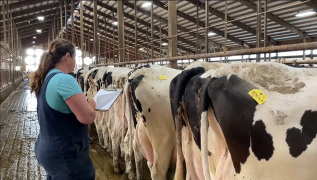
For further examination, cows should be in the headlocks for one hour or less to minimize the amount of time away from other behavioral needs and maximize milk production. To limit the time cows are in headlocks for examination, come prepared for the fresh cow exam.
- Do you have a list or report of all fresh cows, with fresh dates, and milk production?
- Is your fresh cow cart or area clean and well organized and stocked with the appropriate treatments needed for the number of cows to be examined?
- Do you have all the tools needed to implement the treatment protocols?
- Is all the equipment being used in good repair?
- Do you have a system to clean and disinfect equipment between cows or uses?
Which cows get examined
It is stressful for fresh cows to be confined or held in headlocks for a long period of time. We can limit the number of cows getting a full examination to those that have a specific reason to examine in more detail. Included on the shortlist are cows that had:
- Calving difficulties
- Twins
- Lame or walking abnormally
- Milk weight deviations
- Udder not full before milking, abnormal udder or quarter
- Abnormal appetite
- Abnormal manure (diarrhea or stiff manure)
- Foul-smelling uterine discharge
- Poor rumen fill
The physical cow exam
The goals of a physical exam are to identify sick cows early, treat sick cows early, prevent the spread of disease, protect the food supply, and improve animal welfare.
Temperature:
Taking temperatures is a key component in the daily monitoring of cows for fever. It is one of the first signs of an underlying infection. A cow’s normal temperature is between 101.50 to 102.50 F. A temperature over 103.0F, or 1.50F above the fresh cow group average during heat stress, may indicate a cow is fighting off an infection or inflammation.
Dehydration:
Not as accurate as observing the cow for sunken eyes, one can perform a skin tent test to determine the severity of dehydration. To conduct the skin test, pinch some skin at the neck, twist it about 900, and let it go. In a sufficiently hydrated cow, the skin should snap right back in place, while in dehydrated cows the skin returns much more slowly. If the skin tent is slightly prolonged for 2 to 4 seconds or prolonged for 4 to 8 seconds, she is considered mildly or moderately dehydrated, respectively. If the skin tent remains tented and does not snap back into place, she is severely dehydrated.
Retained placenta (fetal membranes):
Retained placenta in cows is defined as a failure to expel fetal membranes within 24 hours after calving and normal expulsion occurs within 3 to 8 hours after calf delivery. Retained placentas are identified by varying amounts of degenerating, discolored, placenta protruding from the vulva for 24 hours or more after calving. Occasionally, the retained placenta may remain within the uterus, which then is signaled by a foul-smelling discharge. In most cases, there are no clinical signs of systemic illness. If there are clinical signs such as fever, it is generally related to toxemia.
Uterine infection (endometritis, pyometra, metritis):
Endometritis is a common mild infection of the uterus, affecting up to 40% of cows
post-calving. The uterus contains pus and the cow may have pus-like discharge from the vagina. The cows do not seem sick and will still eat.
Pyometra is another type of uterine infection that occurs when the cervix closes and pus is trapped in the uterus, stopping the cow from cycling.
Metritis is a severe infection of the uterus that can cause sick cows, a drop in milk production, fever, and even death. It is characterized by the presence of an abnormally enlarged uterus, a foul-smelling watery, red-brownish discharge with systemic illness and fever.
Diagnosis of a uterine infection requires a veterinarian to examine the reproductive tract for pus, 7 to 28 days after calving. At-risk cows, those who have reduced uterine health from twins, difficult birth, uterine tear, dead calf, or retained placenta, and/or those who have visible discharge should be examined.
Mastitis:
Mastitis is an infection within the udder and fresh cows are at increased risk for mastitis. They are most susceptible to infection during the first few weeks at dry off and right at calving. The simplest approach to identifying mastitis is monitoring for clinical mastitis by stripping the cow’s udder looking for abnormal milk including clots or flakes and subclinical mastitis by using a CMT paddle to determine if cows are over 200,000 SCC, an indication of infection. Sterile milk samples can be collected to determine the type of pathogen for future treatment decisions.
The cow’s metabolic status
Metabolic status is just as important as infectious disease. Many metabolic disorders can be secondary to infection as cows may go off feed when sick, negatively impacting the cow’s energy and nutrient status which can lead to metabolic disorders.
Ketosis:
Ketosis is the most common metabolic disorder in high-producing dairy cows during the first 6 to 8 weeks of lactation. Since it tends to be a common concurrent disease with DAs, retained placentas or fetal membranes, and metritis we can look at it as a symptom of another problem that warrants further examination. Ketosis occurs when the body burns fat for energy instead of glucose. This occurs when the cow is off feed and begins to use her fat reserves to supply the much needed energy for maintenance and lactation. Key clinical symptoms include loss of appetite, decreased milk production, noticeable loss of body condition, empty-appearing abdomen, firm dry manure, acetone-like odor from their breath and urine, and in some cases neurological nervous signs; however, ketosis can be subclinical with no visible outward signs.
To diagnose ketosis in cattle, a common cow-side measurement of ketone bodies is the use of a strip test that measures acetoacetate and acetone concentrations in urine with reasonably accurate interpretation within 5 to 10 seconds. The strip test is read by observation of a particular color change which represents a specific level of ketone bodies in the urine. However, the most accurate cow-side method for identifying ketosis is measuring the levels of one of the primary ketones in the blood, β-hydroxybutyrate (βHBA), with a meter.
Milk Fever:
Milk fever is a metabolic disorder caused by insufficient blood calcium levels, commonly occurring around calving. Most cases occur between calving and approximately 3 days after calving. Milk fever is divided into 3 stages based on clinical signs.
Milk fever can lead to many other metabolic problems in cows, including retained placenta, ketosis, mastitis, uterine infections, and fatty liver syndrome.
Stages of Milk Fever
| Stage 1< 1 hour | Stage 21 to 12 hours | Stage 3 | |
| Clinical signs | Loss of appetite Excitability Nervousness Hypersensitivity Weakness Weight shifting Shuffling of hind feet | Turn head into flank Dull Listless Cold ears Dry nose Incoordination with walking Muscle trembling Inactive digestive system Constipation Decreased body temperature Rapid heart beat | Inability to stand Progressive loss of consciousness Death |
Displaced abomasum (DA):
The abomasum is the fourth, or true stomach, of the cow and is located on the floor of the abdomen in the right front quadrant. Because the abomasum is suspended loosely in the abdominal cavity, it can occasionally float and be displaced to the left, becoming trapped between the rumen and the left abdominal wall. This can occur when its muscular wall loses tone and it becomes partially gas-distended. When in this position, the entrance and exit to this stomach become slightly kinked, reducing the passage of food and causing the accumulation of fluid and gas. This results in dullness in cows, reduced milk production, and firm manure.
Uncomplicated ketosis, retained placentas, metritis, and milk fever at calving are risk factors for DA. Assess rumen activity by placing the fist in the paralumbar fossa, or the triangular area between the last rib, under the loin, and in front of the hip, feeling for rumen contractions, which should occur once or twice per minute.
Listen for abnormal sounds that are produced when the abomasum becomes trapped high on the left side and is enlarged with gas. In the same area, you observe for rumen fill, thump the cow listening for a “ping” indicating a DA. A DA will produce a distinct high-pitched “ping” from the thumping sounds bouncing back from the gas-filled abomasum and sound similar to a penny falling in a well or an over-inflated basketball.
Conclusion
Fresh cow health is one of the most important aspects of a dairy operation. Starting a cow on the right foot is imperative for a successful lactation where all cows are meeting or exceeding their full potential. Examining cows daily during their fresh period will enable farmers to notice subtle changes in a cow’s behavior to minimize the impact of fresh cow diseases and metabolic disorders.

 From birth to strong steps: Understanding and managing knuckle over pasturns in calves
 🎧 Listen: Lameness affects up to 50% of dairy cows during their lifetime
 Feeding Zeolite A for Milk Fever Prevention in Dairy Cattle
 How Lung Ultrasounds Are Changing Calf Care


