Introduction
Bovine respiratory disease (BRD) remains one of the most common and costly health challenges in preweaned dairy calves. While traditional diagnosis relies on visual symptoms, such as coughing, nasal discharge, or elevated temperature, these signs often appear late in the disease process, after damage has already occurred. Even when calves appear healthy, subclinical lung infections — or non-visible forms of pneumonia — can compromise growth, feed efficiency, and future milk production.
According to Dr. Terri Ollivett from the University of Wisconsin School of Veterinary Medicine, the lungs can be considered an ‘indicator organ’ because respiratory disease often reflects broader management issues, such as poor passive transfer of immunity, poor nutrition, or environmental stressors. By using lung ultrasonography, producers and veterinarians can detect disease sooner and make better decisions to improve calf health.
Early Detection of Subclinical Pneumonia
Ultrasound has been a valuable tool for revealing what the eye cannot see. In recent years, farmers and veterinarians have used lung ultrasound to visualize a calf’s lungs in real time and detect subtle signs of respiratory disease. Dr. Ollivett says that, depending on the farm, 50-80% of pneumonia cases are present subclinically for 7-14 days before clinical signs are observed. By identifying these cases early, farms can intervene with targeted treatment and management adjustments before long-term health and growth are compromised.
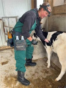
Dr. Kendra Wells of Valley Veterinary Clinic routinely conducts calf lung ultrasounds at Wisconsin dairies. She explained that ultrasound data can be a valuable management tool, noting, “Sometimes calves with severe pneumonia don’t show clinical signs, but the ultrasound reveals the disease.” (Image 1)

Studies have shown that calves with lung lesions detected by ultrasound, even if they do not exhibit any clinical signs of illness, still exhibit impaired growth and delayed development. A 2017 study of more than 600 Holstein heifers found that those with lung consolidation (LC) detected at weaning were less likely to become pregnant and more likely to be removed before first calving than heifers without consolidation (NC). A 2018 study of 215 Holstein calves found that having ≥ 3cm of lung consolidation during the first 8 weeks of life resulted in a 1155 lb decrease in first-lactation 305-d milk production.
Calf Lung Ultrasound Scoring System
Lung ultrasound in dairy calves is a practical method for assessing lung health and is particularly valuable for farms experiencing a high prevalence of subclinical pneumonia. (Image 2) The person conducting the ultrasound looks for a series of indicators when determining lung scores.
Normal Lung
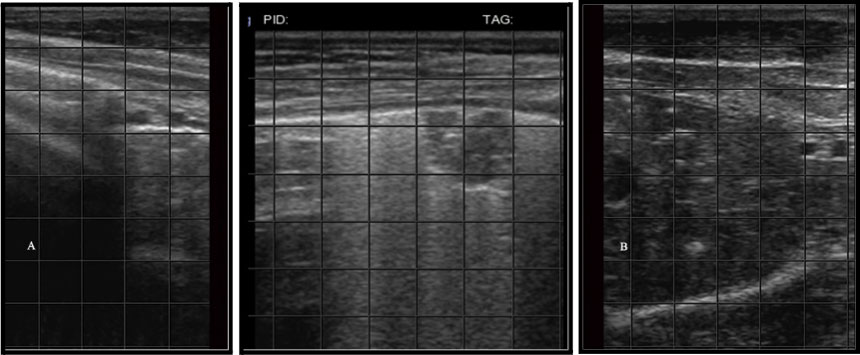
A normal lung is air-filled and appears on the ultrasound image as a series of horizontal white lines (called reverberation artifact) that move back and forth as the calf breathes. The most superficial of those horizontal lines, the pleural line, represents the boundary between the chest wall and the surface of the lung. A smooth, continuous pleural line indicates healthy lung tissue.
Comet Tails
Comet tails are vertical, bright white lines that emanate down from the surface of the lung. Normal lungs may have a few comet tails, but the presence of severe, diffuse comet tailing is suggestive of interstitial disease due to increased fluid or cellular accumulation within the lung tissue. Interstitial disease can result from septicemia, viral infection, or pulmonary edema from heart failure, fluid overload, or anaphylaxis.
Lung Consolidation
Lung consolidation is a condition in which part of the lung has lost its normal air content and become filled with inflammatory debris. Ultrasound images will show consolidated areas as solid, gray areas that lack the white horizontal lines typical of reverberation artifact. The severity score is determined by the size and extent of consolidation (Table 1).
Pleural Effusion, Abscesses, Necrosis
Pleural effusions (fluid accumulation between the lung and the chest wall), abscesses (round, encapsulated fluid and gas-filled pockets), and necrosis (solid non-encapsulated pockets within consolidated lung) are three other lesions that can be detected on lung ultrasound. Pleural effusions have multiple causes and should be interpreted in conjunction with a thorough physical examination of the calf. Abscesses and necrosis indicate chronic lung disease, which often carries a poor prognosis.
The lung scoring system typically ranges from 0 to 5 based on the appearance of lung tissue in the ultrasound image (Table 1). To properly score, the operator must recognize the difference between air-filled lung, air-filled lung with diffuse pleural roughening (also called comet-tail artifacts), lobular lung lesions, and lobar lung lesions. In the context of this US scoring system, lobular and lobar lesions simply reflect the extent to which the lung lobe is consolidated on the ultrasound image. Lobular lesions are relatively small discreet areas of consolidation within an otherwise aerated lung lobe. In other words, the bright white pleural interface with reverberation artifact of normal lung can be seen both above and below the lobular lesion when the probe is placed vertically within the rib space. Lobar lesions indicate full thickness consolidation of the lung lobe that extends proximally from the tip of the lobe. In the US image, the entire distal lung lobe is visible, with the gray parenchyma, and the aerated lung cannot be seen ventral to the lesion (Image 3).
Calves with a score of 2 or higher are generally considered candidates for treatment. This scoring system is valuable for detecting disease before clinical signs, such as coughing or nasal discharge, become apparent, allowing for earlier intervention and improved long-term outcomes. As a reminder, treatment is indicated in calves with clinical signs of respiratory disease even if their lungs appear normal on ultrasound.
Table 1.
| Score | Ultrasound Findings | Interpretation |
|---|---|---|
| 0 | Normal lung: bright white pleural line, few to no comet tails, no consolidation | Healthy lung |
| 1 | Severe, diffuse comet tail artifacts (B-lines all emanating from a bright white pleural line) | Interstitial disease |
| 2 | Lobular consolidation ≥ 1 cm | Mild lung consolidation |
| 3 | Lobar consolidation in 1 lobe | Moderate lung consolidation |
| 4 | Lobar consolidation in 2 lobes | Moderate to severe lung consolidation |
| 5 | Lobar consolidation in 3 or more lobes | Severe lung consolidation |
Improved Treatment Outcomes
When respiratory disease is detected early, treatment is more likely to be effective. Calves with mild lung lesions identified by ultrasound tend to respond more effectively to antimicrobial treatment and supportive care when housed in clean, dry, and draft-free environments that support recovery and help prevent long-term lung damage. Lung ultrasound also provides a means to monitor healing over time, helping to avoid unnecessary retreatment and reduce overall antibiotic use, which is a win for both calf health and antimicrobial stewardship.
Better Decision-Making for Management
Ultrasound findings can guide important management decisions on the farm. By identifying the severity of lung damage, producers and veterinarians can make more informed choices about whether to remove the animal from the herd or retain individual calves. Some farms also use lung scores to adjust the timing of weaning or regrouping, giving vulnerable calves extra time to recover and improving their chances of a successful transition.
Benchmarking and Herd-Level Insight
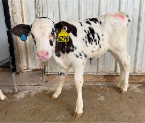
When used regularly, lung ultrasound can serve as a benchmarking tool, allowing producers to evaluate the effectiveness of colostrum programs, ventilation, sanitation, and vaccination strategies. Herd-level lung health trends can highlight areas for management improvement and support continuous progress in calf-rearing practices. (Image 4)
Practical Implementation and Cost
While ultrasound equipment requires an initial investment for the veterinary clinic, portable and user-friendly devices are increasingly accessible for on-farm use. With proper training, veterinarians can scan a calf’s lungs in less than a minute. The value lies in earlier and more accurate detection of pneumonia, which can lead to better management, lower mortality, improved calf growth, and reduced treatment needs.
Incorporating veterinary expertise offers a practical way to utilize this tool. Lung ultrasound detects more cases of respiratory disease than clinical scoring alone, uncovering subclinical problems that might otherwise remain hidden. Although economic returns vary with herd size and management, research consistently shows that calves with severe lung lesions gain less weight per day and produce less milk in the future than their healthier counterparts.
Conclusion
Lung ultrasonography is transforming the way we understand and manage calf respiratory disease. By looking beyond visible symptoms, this technology equips farms with the insights needed to make proactive, data-driven decisions that support long-term calf health, productivity, and welfare. In the ever-evolving landscape of dairy management, lung ultrasound isn’t just a diagnostic tool—it’s a strategic advantage.
Author

Aerica Bjurstrom
Regional Dairy Educator – Aerica’s work focuses on herd health and animal welfare. She also has a strong background in meat quality and has done programming in market cow carcass quality.

Angie Ulness
Dairy Educator – Angie has been active in the dairy industry her entire life. She was raised on her family farm in Door County and currently farms with her husband Mark and their four children in Manitowoc County. Angie has previously worked as a Field Representative for Holstein USA and a Senior Territory Manager for a Pharmaceutical company. Her area of focus is Dairy Farm Management, Profitability and Efficiency.
Published: February 11, 2026
Reviewed by:
- Jennifer Van Os, Dairy Animal Welfare Extension Specialist and Associate Professor at the University of Wisconsin–Madison
- Theresa Ollivett, Associate Professor in the Food Animal Production Medicine at the University of Wisconsin–Madison
References
- Balchem. (2024, Nov 5). Keep them breathing easy: Diagnosing calf respiratory problems with ultrasound [Webinar, Episode 121]. Real Science Lecture Series. Balchem. https://balchem.com/anh/podcasts-webinars/diagnosing-calf-respiratory-problems-with-ultrasound/
- Cramer, M. C. (2018). Determining differences in growth, behavior, and serotonin in dairy calves affected by respiratory disease as diagnosed by lung ultrasound and a clinical respiratory score (Doctoral dissertation, University of Wisconsin–Madison). University of Wisconsin–Madison Institutional Repository. https://asset.library.wisc.edu/1711.dl/V6VAB4WDGSAHT8Y/R/file-aa558.pdf
- Cramer, M. C., & Ollivett, T. L. (2019). Growth of preweaned, group-housed dairy calves diagnosed with respiratory disease using clinical respiratory scoring and thoracic ultrasound-A cohort study. Journal of Dairy Science 102:4322-4331. https://doi.org/10.3168/jds.2018-15420.
- Dunn, T. R., Ollivett, T. L., Renaud, D. L., Leslie, K. E., LeBlanc, S. J., Duffield, T. F., and Kelton, D.F. (2018). The effect of lung consolidation, as determined by ultrasonography, on first lactation milk production in Holstein dairy calves. J. Dairy Sci. 101:5404 – 5410. https://doi.org/10.3168/jds.2017-13870.
- Ollivett, T. L., & Buczinski, S. (2016). On-farm use of ultrasonography for bovine respiratory disease. Veterinary Clinics of North America: Food Animal Practice, 32(1), 19-35. https://doi.org/10.1016/j.cvfa.2015.09.001
- Taio, G., Lisuzzo, A., Cecchini, F., Tommasoni, C., Gianesella, M., & Fiore, E. (2025). Field application of lung ultrasonography in bovine: A scoping review. Large Animal Review, 31(1), 53–60. University of Padua, Department of Animal Medicine, Production and Health (MAPS). https://www.largeanimalreview.com/index.php/lar/article/view/850/307
- Teixeira, A. G. V., McArt, J. A. A., & Bicalho, R. C. (2017). Thoracic ultrasound assessment of lung consolidation at weaning in Holstein dairy heifers: Reproductive performance and survival. Journal of Dairy Science, 100(4), 2985-2991. https://doi.org/10.3168/jds.2016-12016.